Nora Eccles Harrison Cardiovascular Research & Training Institute

Effects of Epigenetics in Cardiovascular Disease & Research

Effects of Epigenetics in Cardiovascular Disease & Research

Strand of DNA on a Blue Background
DNA methyl transferase I (mid) transfers a methyl group from S-adenosyl methionine (red) mostly to cytosine bases of the human DNA.

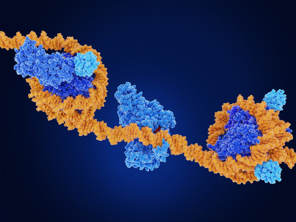
In biology, epigenetics is the study of heritable changes that do not involve alterations in the DNA sequence. The Greek prefix ‘epi’ means over, outside of, or around. Thus, its definition is the feature of the genetic code that exists or develops in addition to traditional genetic inheritance.

Epigenetics is, in essence, the study of how things like lifestyle choices and the environment can cause changes that affect the way your genes work, including what diseases you become predisposed to. Most epigenetic changes are reversible; however, their effects can worsen or cause diseases that are not.

Epigenetics has emerged as a powerful tool to aid in the therapeutic treatment of cardiovascular disease by looking closely at the epigenetics of cardiovascular patients and customizing treatment plans based on someone’s unique epigenetic factors.

Studies That Link Epigenetics and Cardiovascular Disease

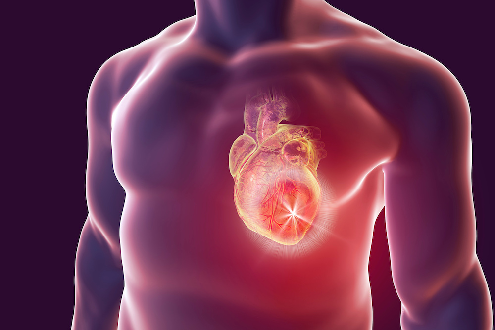
Epigenetics was initially studied in patients with cardiovascular disease to better understand their prominent role in inflammation and vascular involvement. These studies in cardiovascular patients showed a significant number of certain epigenetic modifications affecting the development and progression of cardiovascular disease.

In the case of cardiovascular hypertrophy, epigenetics studies found that cardiac hypertrophy is linked with histone acetylation, implicating both histone acetyltransferases (HATs) and histone deacetylases (HDACs).
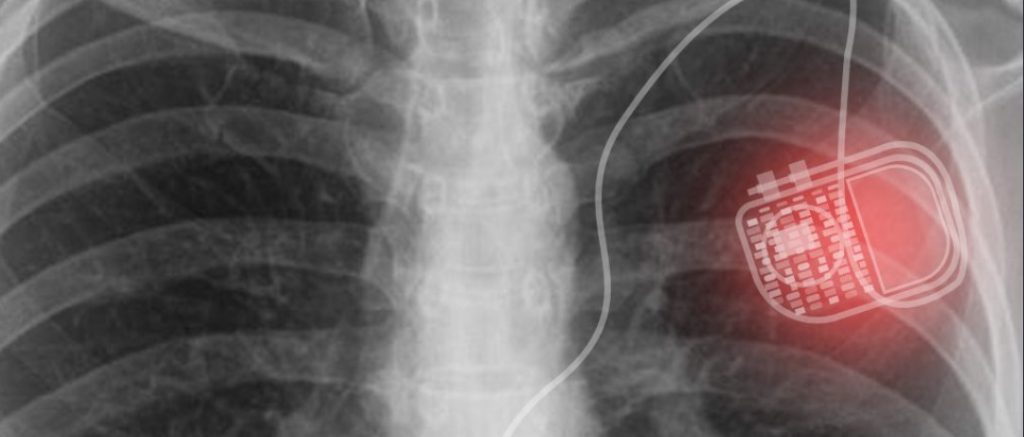
Cardiac Arrhythmias

Epigenetics has also been studied in afflictions like cardiac arrhythmias. Atrial fibrillation is one of the most common heart arrhythmias, particularly in older patients, and can be deadly. In a study done on its role in atrial fibrillation, transgenic mice were programmed to develop cardiac hypertrophy, and then treated with an epigenetic approach, “Liu and colleagues showed that an injection of a specific HDAC inhibitor reverses atrial fibrosis and diminishes atrial fibrillation vulnerability following an electrical stimulation.” (1)

Among miRNAs that have been widely studied in experimental environments, miR-1 was found to be essential in normal electrophysiological conduction. When miR-1 was deleted in rodents, the rate of sudden death was much higher. This points to the idea that the miR-1 subtype of miRNAs is essential to healthy cardiac rhythms and lowering levels of miR-1 could signal an oncoming arrhythmia. Epigenetic interventions can more effectively return the heart back to a normal state when combined with traditional methods.

Atherosclerosis

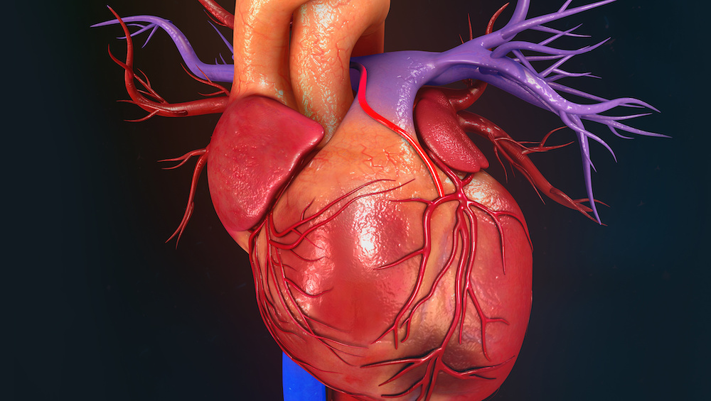
In atherosclerosis (hardening of the arteries or artery disease), epigenetic studies reveal some key factors to recognize developing atherosclerosis, sometimes before the patient even feels symptoms. DNA methylation, miRNAs, and epigenetic mechanisms have all been described in atherosclerosis. Two studies were done that show coexisting DNA methylation alongside atherosclerotic lesions in rodents. Interestingly, it was observed that DNA methylation could often be detected in animals far before the anatomical presentation of the disease itself. In short – DNA methylation testing could stand as a powerful screening tool to determine the early stages of atherosclerosis.

Diabetes and Cardiovascular Disease

In studying the relationship between diabetes and cardiovascular disease, epigenetics can be used to demonstrate the effect of untreated or poorly treated diabetes on worsening heart disease. In a study conducted and published by Louisa Villeneuve and colleagues, they found connections between diabetes and the inflammatory epigenetic reactions that can be caused by high glucose.

This study found that the occurrence of inflammatory genes in vascular cells that were exposed to high glucose for extended periods was increased. (2) Conversely, they found that histone H3 (specifically, H3K9me3), which is known to guard the body against the biochemical effects of diabetic inflammation, were decreased in environments in which they had to succumb to high glucose. The finding shows that uncontrolled glucose levels (usually caused by uncontrolled or poorly treated diabetes) can simultaneously cause inflammatory reactions while hindering the effectiveness of key histones like H3K9me3.

In all studies that have been completed, one thing was clear. Epigenetic changes are a clear indicator that can tell us important information about a patient’s ongoing cardiovascular disease or likelihood of developing impending cardiovascular disease.

Conclusion on Epigenetics and Cardiovascular Research

Epigenetics are genetic changes that happen in reaction to external factors. These external factors are usually caused by environmental or lifestyle exposures. Epigenetic changes can be important ‘warning signs that a disease is worsening or about to occur. Furthermore, by studying epigenetics in relation to heart disease, we can get a better idea of what prevention and intervention tactics should be taken.

Epigenetic factors and expressions are somewhat unique and can vary from patient to patient, but overall, there are quantifiable patterns of what role epigenetics plays in cardiovascular disease. Studying these micro-changes can make a big difference in preserving the quality of life in patients with cardiovascular disease and aid in early detection of patients with epigenetic risk factors for the disease.

Cardiovascular Research and Training Institute

Researchers at the Nora Eccles Harrison Cardiovascular Research and Training Institute (CVRTI) are focused on understanding the epigenetic factors that alter transcription and thereby cardiac physiology during the development of arrhythmias, cardiac hypertrophy, and heart failure. Specifically, research at CVRTI has centered on understanding the role of lysine methyltransferases in modifying histones within the nucleosome, characteriz ing specific histone modifications that are differentially regulated during disease, and identifying the role of transcription factors  in pathological remodeling and reverse remodeling . These studies have utilized zebrafish , mouse models and human induced pluripotent stem cells  as transgenic models of disease to further our understanding of these key processes. By incorporating cutting-edge technology with novel animal models of disease, the epigenetics research at CVRTI is making significant contributions not only to basic science, by elucidating the fundamental basis of gene expression and transcriptional regulation in the cardiomyocyte, but also to the clinical realm by identifying key regulators of cardiac pathophysiology that may also protect the heart from ischemic injury or heart failure.    

What is the Difference Between Pacemakers and Defibrillators?

What is the Difference Between Pacemakers and Defibrillators?

Closeup Image of Pacemaker in Chest

The heart is the most important muscle in the body—it’s the one that keeps us alive. When the heart can’t keep up due to disease, damage, or other issues, technology is available to help. Two of the most common devices associated with heart attacks, heart disease, and other heart conditions are pacemakers and defibrillators. But what are these tools, and what makes them different? How does each provide the assistance that the heart needs to continue beating (since that’s what both were designed for)?

In this blog, we’ll take a look at defibrillators and pacemakers to explain when and how they’re most commonly used and what types of patients are candidates for treatment with either device.

Defibrillators

Also known as ICDs, implantable cardioverter defibrillators are implanted devices that are designed to shock the heart to assist in restoring its rhythm to normal. They are usually prescribed for those who at risk of having life threatening arrhythmias. These devices can detect the dangerous arrhythmias and convert the rhythm back to normal using electric shock.

Anyone who is at risk from arrhythmia-induced death can benefit from an ICD, including people with cardiomyopathy, a history of cardiac arrest, severely reduced heart function, or genetic diseases like Long QT syndrome, ARVC, cardiac sarcoidosis, etc. that can cause ventricular arrhythmias (fast rhythm coming from the ventricles or the bottom chambers of the heart).

ICDs are usually implanted below the collar bone. An ICD is larger than a pacemaker and about the size of a pager. The defibrillators are used for tachycardia or fast heartbeats, but these devices can also provide dual function and serve as a pacemaker to correct issues with slow or weaker heartbeats, too.

Defibrillators include the generator along with the leads that are used to detect rhythm and deliver the appropriate shock to the heart as needed. The generator goes below the collar bone, and it houses the computer, capacitor, and the battery. The lead are wires that run through a large vein that goes to the heart so that they can deliver the shock needed to restore the rhythm. These devices offer a longer lifespan in general. For some patients they also can offer a better quality of life and longer lifespan but those do require slightly more invasive surgical methods.

Risk of procedure include infection, bleeding, and the potential for unnecessary shocks. For the most part, risks are minimal, and most people see more improvement than anything.

Pacemakers

The pacemaker is also an implanted battery-operated device, smaller than the ICDs, that assists the heart in beating at an acceptable rate. It is used for people who have a slower heart rate.

Implanting a pacemaker requires a minor surgery and a short hospital stay just like the ICDs. They can improve people’s quality of life in several ways by providing the electrical impulses the heart needs to keep the heart beating and can’t produce on its own.

The pacemaker is also implanted below the collar bone and features leads and a generator. The generator is where the information and battery are held, and the lead wires are run through a large vein that goes to the heart so that they can deliver the impulses that give the heart the reminder to beat. There are newer pacemakers available now that don’t have a lead and are referred to as “leadless” pacemakers. Leadless pacemakers are implanted directly in the heart itself and function as a complete unit.

In rare instances, infection or other complications can occur, but they are rare. Some people also worry about the interference of electronics or magnets, but again, that’s a minimal risk for the most part.

Conclusion

Ultimately, patients are best served by discussing their condition with their cardiologists to determine the right solution. Understanding the differences between these devices and the conditions they treat can help you make the most informed decision about keeping your heart (or your loved one’s) healthy and beating strong for as long as possible.

These electric devices are constantly evolving and improving, so it will likely only be a matter of time before there are even better solutions available. The heart is a strong muscle, typically, but even it is not impervious to damage and ill health as time goes on. For those with heartbeat irregularities, a pacemaker or defibrillator implant could be the solution they need to provide a better quality of life and give them a better chance of living longer.

Cardiovascular Research and Training Institute

At CVRTI, we are investigating multiple aspects related to pacing and defibrillation. We are actively investigating more efficient ways  to pace patients out of dangerous arrhythmias using the highly efficient conduction system in the heart. This approach can potentially lower the chance of getting shocked, which is uncomfortable for patients. There is also active investigation exploring many different pathways to improve muscle function in heart failure  using gene therapy based medicine . These novel therapeutics will lower the risk of developing dangerous cardiac arrhythmias. There is also more basic science investigation exploring genetic and molecular basis of developing heart failure and arrhythmias, with the development of intravenous therapies to stop dangerous arrhythmias as they occur.


What is Myocardial Recovery?

What Is Myocardial Recovery?

Red Transparent Body with Heart - What Is Myocardial Recovery Blog Graphic

Heart failure (HF) is characterized by a pathologic process known as “remodeling” that involves impairment of the function of the heart and progressive dilation of the chambers of the heart. The process of remodeling is associated with adverse cellular, structural, and functional myocardial changes, that had long been deemed progressive and unidirectional. Clinical experience has shown that the process of remodeling can be delayed or even reversed, either spontaneously in the setting of acute cardiac injury (e.g., acute myocarditis and other), or to be facilitated through guideline-directed HF medical therapy including cardiac resynchronization therapy in chronic HF . Mechanical circulatory support (MCS) with left ventricular assist devices (LVADs) is an established treatment modality for patients with advanced disease. Besides their role in supporting the systemic circulation (more info provided below), they provide significant off-loading of the heart, creating a favorable environment for reversal of the structural and functional alterations of the failing heart, a process known as “reverse remodeling” . It has been repeatedly shown that a subset of advanced HF patients can significantly improve their cardiac structure and function while on durable LVAD, to the point where withdrawal of the LVAD support can be considered . In light of these findings, the concept of HF irreversibility has been refuted and the notion that severe HF indicates irreversible end-stage disease has been revised. The National Institutes of Healtdddh (NIH) organized a working group of experts from around the globe and the consensus recommendations derived from this body defined myocardial recovery as a reversal of the pathological state of the myocardium with significant improvement in heart structure and function sufficient to achieve a sustained remission from hospitalizations and other adverse events that take place in patients with chronic heart failure.

Left Ventricular Assist Device (LVAD)

A left ventricular assist device is a form of mechanical circulatory support used in the treatment of patients who have been diagnosed with advanced HF. An LVAD is a pump that helps your heart circulate blood from the left ventricle to the rest of the body. LVAD devices are implantable devices commonly used as a bridge to transplant in patients for whom medication alone didn’t improve their heart function. They are also used as lifetime permanent therapy in advanced HF patients that are not eligible for heart transplantation due to various contraindications.

Studies have shown that using an LVAD in combination with guideline directed medical therapies  have the potential for increasing the probability for recovery from HF. These studies were conducted using standard drug therapy protocol, optimization of the LVAD speed, and consistent testing of the myocardial function in participants . The ultimate goal is full myocardial recovery with LVAD explantation with no recurrence of heart failure symptoms.

Reverse Remodeling

A main function of the heart is to pump blood to the lungs and the rest of the body. When a patient experiences heart failure, the heart isn’t working correctly, and blood is not pumped adequately from the heart to the rest of the organs. This can lead to a back-up of fluid in the lungs as well as a decrease in the amount of blood the heart ejects. Some patients who experience heart failure require a transplant or the assistance of an LVAD.

Reverse remodeling represents a significant improvement of the heart due to a positive response to treatment . When reverse remodeling occurs, there is an improvement in the heart’s ability to fill and eject blood to the body. Doctors often notice improvements to the size of the ventricle. As the ventricle size normalizes, it often leads to improved heart function (ejection fraction is one of the metrics of heart function that is widely used). When reverse remodeling is identified, doctors can optimize the pump speed of the LVAD and continue with pharmacological treatment to advance the reverse remodeling and facilitate myocardial recovery .

Advancing the Science of Reverse Remodeling and Heart Recovery

Understanding the biological mechanisms driving heart recovery following LVAD may be paramount to understand and facilitate cardiac improvement in the broader HF population. By examining heart tissue samples from patients with various degrees of LVAD-mediated cardiac improvement could lead to the discovery of novel therapeutic targets . The overarching goal is to improve our understanding of cardiac biology and the associated molecular, cellular, and structural changes that are implicated in cardiac recovery, and manipulate them in a way that could be beneficial for the greater HF population. This research is currently being conducted by several investigators at the Nora Eccles Harrison CVRTI  and the University of Utah Health (visit lab websites listed below in the corresponding section).

Conclusion

Heart failure is a progressive disease in which the heart does not function properly to pump blood throughout the body and medications are only one way to treat heart failure. When patients don’t seem to respond to pharmaceutical treatments, they may undergo mechanical circulatory support with a device known as an LVAD. The LVAD is implanted into the left ventricle to improve pumping of blood from the heart to the rest of the body. Studies show that if standard medical therapy is used in conjunction with the LVAD, the heart can begin to regain function and show reverse remodeling. Some patients are fortunate enough to experience significant heart reverse remodeling and sustainable myocardial recovery.

CVRTI Labs involved in HF and Myocardial Recovery Research:

What Is Heart Failure Therapy?

What Is Heart Failure Therapy? 6 Potential Heart Failure Treatments

Full Color Graphic of Heart Close Up

Cardiovascular disease resulting in heart failure is the leading cause of death for people living in developed countries. In an effort to find better ways to treat CVD, scientists have been researching a variety of heart failure therapies. Some of those are discussed in this article.

Omecamtiv Mecarbil

Omecamtiv mecarbil was developed as a safer alternative to inotropic agents while still having similar pharmacological effects. It specifically acts as a cardiac myosin activator. It binds to myosin and causes a shift in the equilibrium of ATP hydrolysis during a stroke.

Studies of omecamtiv mecarbil show that it was well tolerated by patients. Phase two trials had promising results as well, so expectations are high for phase three trials.

Ularitide

Elevated levels of a family of hormones called natriuretic peptides indicate heart failure. Three main natriuretic peptide receptors exist, NPR-A, NPR-B, and NPR-C. NPRs have been a component in the beneficial treatment of heart failure, so they are targeted for pharmacological therapies.

Ularitide is a synthetic version of urodilatin, a kidney peptide hormone. Ularitide binds with NPR-A, which causes diuresis and natriuresis to increase. Studies of ularitide therapy indicate that it can reduce cardiac wall stress and produce beneficial hemodynamic effects, but it doesn’t affect disease progression.

Serelaxin

The location of the receptors in the body is important in cardiovascular therapy. Serelaxin is a synthetic version of relaxin; the peptides in relaxin have a structure similar to insulin. Currently, four relaxin receptors are known to be present in major body organs. These receptors exist in the kidney, blood cells, heart, and lungs.

Trials of Serelaxin have had mixed results. Tests with primarily Caucasian patients indicated that infusions of Serelaxin were more effective than oral doses. However, similar trials with Asian patients showed no significant improvement or change in mortality rates with Serelaxin therapy.

Tolvaptan

A consequence of heart failure is fluid retention. This fluid retention leads to edemas and pulmonary congestion. During heart failure, arginine vasopressin levels increase, and higher levels are part of the advanced stages of the disease. Sodium retention and neurohumoral abnormalities are the leading causes of fluid retention. 

Loop diuretics are one of the common treatments for heart failure, but this therapy doesn’t work in approximately 30% of patients. Loop diuretics also cause hypotension or electrolyte imbalances in some patients. Tolvaptan is a treatment that is less likely to worsen kidney functions than typical diuretics.

The benefits of using tolvaptan to treat heart failure have been assessed in various studies. Results of one such study showed that symptoms of heart failure were decreased, but some patients developed hypernatremia at increased doses of tolvaptan.

CT-1

CT-1 is connected to multiple heart-related issues. CT-1 is associated with myocardial fibrosis, the stimulation of fibroblasts, and heart valve disease, among others. CT-1 affects the entire cardiac muscle, not just single cardiomyocytes. The pathological changes created by CT-1 are similar to the physiological changes seen in athletes.

The physiological changes are not as beneficial as the ones brought about by CT-1. Another difference is the reversibility of the changes. The CT-1 changes are completely reversible.

Gene Therapy

Some doctors and scientists believe gene therapy is the best hope for rare congenital diseases. However, there hasn’t been a tremendous success with it, so many scientists think the expectations of success are unfounded. There is also hope that gene therapy could be of great benefit to the field of cardiology.

The main targets of gene therapy are proteins that regulate calcium levels. This is because people with CV (chronic venous) insufficiency often have imbalances when it comes to calcium regulation within the cardiomyocytes within their hearts. There are two main problems of gene therapy for cardiovascular diseases. They are the reaction of the patient’s immune system to the treatment and the effect of the treatment getting worse over a long time.

While there have been problems with gene therapy, scientists are still trying to improve the techniques. The continued gene therapy testing is a sign that the hope for its success continues to be justified.

Conclusion

Cardiovascular disease is the number one killer of people in developed countries. Scientists have been increasingly working on therapies for heart failure patients due to the increased number of people who suffer from cardiovascular disease. Continued progress means they have hope of finding a therapeutic solution for treating heart failure. All of the therapies discussed in this article show promise for success; however, they need continued research before they are deemed effective treatments.

Cardiovascular Research & Training Institute’s Heart Failure Research

Cardiovascular disease resulting in heart failure is the leading cause of death for people living in developed countries. In an effort to find better ways to treat CVD, scientists have been researching a variety of heart failure therapies. At the Nora Eccles Harrison Cardiovascular Research and Training Institute (CVRTI), researchers are interested in how cardiac muscle biology relates to the mechanistic basis of heart failure(HF). The CVRTI is committed to focusing on learning what drives heart muscle failure and how best to treat it. 

Our Heart Failure Research in the Areas of Gene Therapy and Therapeutic Devices

Researchers are working on changing the standard of care for HF patients through drug therapy, gene therapy and therapeutic devices. Specifically, CVRTI investigators are trying to understand how the failing heart can recover in order to improve outcomes for HF patients. Our investigators are focused on identifying molecular and metabolic targets to develop new therapeutic tools for myocardial recovery. They also evaluate myocardial ultrastructure, microstructure, and function in normal, diseased, and aged hearts. Preserving and optimizing the cellular cytoskeleton and the mitochondrial function in the failing heart are major research interests of our investigators. Specifically, our investigators define the mechanisms by which the cytoskeleton delivers ion channels to their respective subregions on the cardiac membrane, and how delivery is changed during heart failure. 

Targeted Delivery 

This work defined the paradigm of ‘targeted delivery’ which describes how the cytoskeleton delivers membrane proteins directly to their functional membrane subdomain and why there is less delivery in failing heart.  Our CVRTI models involve exploration of Connexin43 gap junction trafficking to the cardiomyocyte intercalated disc and L-type calcium channel trafficking to cardiomyocyte T-tubules. In the process we identified that the mRNA of Cx43 is alternatively translated to generate endogenously up to six different truncated isoforms which are essential to trafficking. These isoforms we identified have a different biophysics and biology as Connexin43 and thus function as new proteins with important roles in basic cell biology. 

cBIN1

A major current focus of our investigators is cardiac BIN1 which regulates L-type calcium channel trafficking, T-tubule folds and is also released into blood as T-tubule origin microparticles, and available as a biomarker of muscle remodeling. We have found that not only is cBIN1 important to calcium hemostasis and electrical stability in ventricular cells, but is reduced in acquired heart failure and is turned over into blood in levels that reflect cardiac content.  We have helped developed a cBIN1 score (CS) as a first of its kind biomarker of cardiac muscle health that could be used as a screen for heart failure, prognosticate heart failure outcomes, and prognosticate the occurrence of ventricular arrhythmia. cBIN1 is now also tested by CVRTI investigators as potential gene therapy for chronic heart failure. 

With these research programs in place, the CVRTI is committed to improving how acute and chronic heart failure patients are diagnosed and treated.