
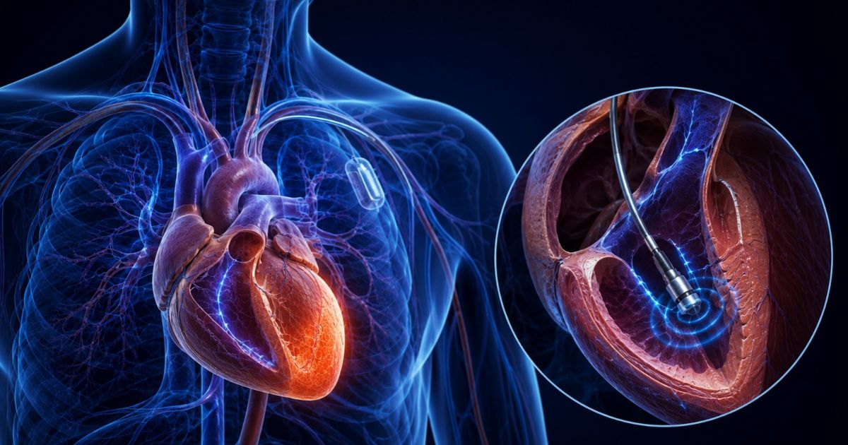
New mapping technologies improve electrophysiology outcomes through higher spatial resolution, structural scar architecture data, and real-time lesion-formation confirmation in medical practice. Many ablation targets are invisible to point-by-point systems, but high-density 3D electroanatomical mapping can help identify the unseen.
CT, MRI, and intracardiac echocardiography with AI integration can reveal midmyocardial and epicardial substrates before catheter placement. Non-invasive electrocardiographic imaging can localize arrhythmia before a procedure. Impedance monitoring can also confirm tissue contact during ablation rather than inferring it.
Let’s explore these developments and examine the clinical evidence to determine where current limitations remain and what improvements may be on the horizon.
How Mapping Technology Guides Catheter Ablation in Cardiology
Catheter ablation delivers energy to the spot generating or sustaining an arrhythmia, so identifying that site is critical for effective care. But an electrophysiologist has to know where the abnormal tissue is located within the cardiac chamber. They also have to determine how the electrical wavefront moves through it and whether the catheter tip makes sufficient contact to deliver energy. One wrong variable can produce an incomplete lesion or procedural complication.
Historically, mapping relied on a small number of contact points recorded sequentially and displayed in two dimensions, with fluoroscopy for spatial reference. For anatomically predictable arrhythmia, that approach was sufficient. But it was insufficient for complex ventricular tachycardia in scarred myocardium. It also didn’t work well for persistent atrial fibrillation or arrhythmias in surgically altered cardiac patients.
New technologies are improving electrophysiology outcomes by changing what the map can tell you. High-density systems can collect thousands of electrograms and render them in three dimensions with temporal activation data layered on top of spatial geometry. Computed tomography and MRI imaging can now be imported directly into the electroanatomical map.
High-Resolution 3D and 4D Mapping Technologies
High-resolution 3D and 4D mapping technologies build chamber geometry using thousands of contact points. These platforms include:
- The Rhythmia HDx (Boston Scientific) — Uses an electrode basket catheter to reduce the time required for adequate chamber coverage in complex substrates.
- CARTO 3 (Biosense Webster) — Combines electromagnetic and impedance-based catheter tracking with voltage and activation mapping to support CT and MRI geometry integration.
- EnSite Precision (Abbott) — Uses an electrode array with beat-to-beat annotation during sinus rhythm and arrhythmia without sustained tachycardia.
High-density technologies are essential because the underlying structures of complex arrhythmias are small, sometimes only millimeters wide. Lower resolution can make narrow isthmuses that sustain macro-reentrant circuits in the atrium easy to miss.
Three-dimensional imaging has also changed what electrophysiologists know about VT circuit architecture, including altered conduction through scar tissue. High resolution can reveal circuits spanning multiple myocardial layers, often with direct procedural implications. Meanwhile, four-dimensional mapping can add time to the spatial model, revealing how activation wavefronts move through the substrate beyond a voltage snapshot.
Single-Catheter Platforms
Ablation workflows generally require switching between mapping and ablation catheters, and each switch introduces the potential for error. But platforms that integrate mapping and ablation into a single catheter let operators map and ablate within the same catheter contacts without withdrawal.
Practical benefits include confirming the substrate within the same catheter delivering energy, removing a source of error and preventing its accumulation while moving between instruments in the same chamber. Talk to your cardiologist today to learn more.
Artificial Intelligence and Multimodal Imaging
Ischemic cardiomyopathy patients may have midmyocardial or epicardial scar tissue that’s invisible to endocardial voltage mapping. So, importing critical structural data from cardiac MRI and CT scans into the electroanatomical map lets teams visualize scar architecture alongside intraprocedurally collected electrophysiological data.
In other words, AI-based scar identification from wall thickness analyses can be integrated with forward-solution ECG mapping. That means non-invasively localizing arrhythmia sources before placing the catheter.
Until recently, imaging consistency across centers and training levels has largely been operator-dependent, creating challenges for health systems seeking consistent procedural workflows. But intracardiac echocardiography provides real-time anatomical visualization that neither MRI nor CT scans can offer.
What This Means for Congenital Heart Disease Patients
Patients with repaired congenital heart disease often have surgical scars, patches, or anatomy that can produce arrhythmia substrates that don’t follow the norm, particularly in a hospital setting where complex cases require specialized mapping. In those cases, dynamic activation mapping is procedurally necessary, not just beneficial.
Noninvasive Electrocardiographic Imaging
Electrocardiographic imaging reconstructs epicardial electrical activation from body surface potentials recorded by multielectrode arrays. This typically includes around 200 to 250 electrodes, each processed against MRI or CT-derived geometries. The output is generally a 3-dimensional map. But technologically advanced platforms, such as VIVO and CardioInsight, help localize arrhythmia sources and identify epicardial exit points for VT circuits.
Noninvasive electrocardiographic imaging also offers preprocedural targeting data that’s useful when the arrhythmia is infrequent or poorly tolerated in lab settings. However, that doesn’t make it a substitute for invasive contact mapping where appropriate. Its primary role is to narrow the target region before introducing the catheter to reduce arrhythmia mapping times.
Reducing Radiation with Electroanatomical Navigation
Early catheter ablation was a fluoroscopy-dependent procedure. But advanced 3D electroanatomical mapping systems have displaced fluoroscopy by using electromagnetic and impedance-based tracking to show the catheter’s position within a virtual display. Some studies even report zero fluoroscopy rates for atrial flutter, SVT, and AF ablation.
Magnetic navigation systems vastly decrease our reliance on fluoroscopic imaging, allowing remote catheter manipulation and keeping the operator out of the radiation field. And since radiation is a clinically relevant variable, especially for operators with high-volume caseloads, reducing cumulative exposure can benefit them and their patients undergoing repeat procedures.
Real-Time Lesion Assessments in Clinical Practice
Delivering radiofrequency energy does not guarantee transmural lesion formation. Other factors, such as catheter stability, local power delivery, tissue contact force, and tissue characteristics, can also determine whether ablation produces electrical isolation or a recoverable injury that reconducts. And pulmonary vein reconnection is one of the best-documented drivers of AF recurrence.
Catheters with contact force sensing can display real-time force between myocardium and the catheter tip during ablation. Meanwhile, dielectric-based tissue characterization tools can help distinguish dense scar, border zone, and viable myocardium at the same tip. This helps complete ablations without relying on procedural voltage mapping thresholds.
How New Mapping Technologies Are Improving Electrophysiology Outcomes
The aggregate effect of new mapping technologies is more information earlier in the procedure, with less reliance on direct inference. High-density maps can identify targets that older, lower-resolution systems cannot. Multimodal imaging integration can also help uncover substrate architectures that intracardiac mapping never could.
For more on electrophysiology research at CVRTI, visit the Nora Eccles Harrison Cardiovascular Research and Training Institute.